Bovine Cryptosporidium Antigen Stool Test Chromatographic Immunoassay
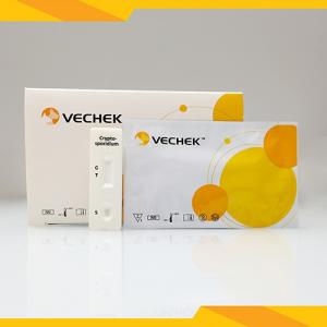
Brand Name:VECHEK
Model Number:VICR-602
Minimum Order Quantity:N/A
Delivery Time:2-4 Weeks
Payment Terms:L/C, T/T
Place of Origin:China
Contact Now
Add to Cart
Verified Supplier
Location:
Hangzhou Zhejiang China
Address:
#383, Qiaoxin Road, Xiasha Street, Qiantang Disctrict, Hangzhou, Zhejiang, P.R.China
Supplier`s last login times:
within 48 hours
Product Details
Company Profile
Product Details
Cryptosporidium Antigen Test 10 minutes Cassette Chromatographic Immunoassay VETERINARY Bovine Range
Product Introduction:
The Cryptosporidium Antigen Rapid Test Cassette is a rapid chromatographic immunoassay for the qualitative detection of Cryptosporidium Antigens in animal feces.
| Production Name: | Cryptosporidium Antigen Test 10 Minutes Cassette Chromatographic Immunoassay VETERINARY Bovine Range | Principle: | Chromatographic Immunoassay |
| Format: | Cassette | Specimen: | Feces |
| Reading Time: | 10 Minutes | Storage Temperature: | 4-30℃ |
| Sensitivity: | 95.50% | Specificity: | 97.70% |
| Accuracy: | 97.50% | Shelf Life: | 2 Years |
Fast results
Easy visually interpretation
Simple operation, no equipment required
High accuracy
What is Cryptosporidium antigen?
Cryptosporidium Antigen, Feces
Cryptosporidium is a parasite that causes cryptosporidiosis, a
profuse, watery diarrhea. Cryptosporidium is the leading cause of
outbreaks of diarrhea linked to water and the third leading cause
of diarrhea associated with animal contact in the world.
Application:
The Entamoeba/Giardia/Crypto Rapid Test Cassette (Feces) is a rapid
chromatographic immunoassay for the qualitative detection of
Entamoeba
histolytica antigens, Giardia lamblia and Cryptosporidium antigens
in human feces.
Description:
Entamoeba histolytica is an anaerobic parasitic amoebozoan, part of
the genus Entamoeba. Predominantly infecting humans and other
primates causing amoebiasis,E. histolytica is estimated to infect
about 50 million people worldwide. Previously, it was thought that
10% of the world population was infected, but these figures predate
the recognition that at least 90% of these infections were due to a
second species, E. dispar. Mammals such as dogs and cats can become
infected transiently, but are not thought to contribute
significantly to transmission. E. histolytica, as its name suggests
(histolytic= tissue destroying), is pathogenic; infection can be
asymptomatic or can lead to amoebic dysentery or amoebic liver
abscess. Giardia lamblia is the most common protozoa known to be
responsible for one of the main causes of severe diarrhoea in
humans, particularly in immunodepressed people. Epidemiological
studies, in 1991, showed that infections with Giardia increased in
the United States with a prevalence of around 6% on 178,000
samples. Generally, the disease passes through a short acute phase
followed by a chronic phase. Infection by G. Lamblia, in the acute
phase, is the cause of watery diarrhoea with principally the
elimination of trophozoites. The feces become normal again, during
the chronic phase, with transient emissions of cysts.
The presence of the parasite on the wall of the duodenal epithelium
is responsible for a malabsorption. The disappearance of
villosities and their
atrophy lead to problems with the digestive process at the level of
the duodenum and the jejunum, followed by weight loss and
dehydration. The majority of infections remain asymptomatic,
however. The diagnosis of G. Lamblia is carried out under
microscopy after flotation on zinc sulphate or by direct or
indirect immunofluorescence, on non-concentrated samples displayed
on a slide. 6 More and more ELISA methods are also now available
for the specific detection of cysts and/or trophozoïtes. Detection
of this parasite in surface or distribution water can be undertaken
by PCR type techniques. The test is based on the detection of a
65-kDA coproantigen, a glycoprotein that is present in the cysts
and trophozoites of G. Lamblia. Cryptosporidiosis is a diarrhoeal
disease caused by microscopic parasites of the genus
Cryptosporidium. Once an animal or person is infected, the parasite
lives in the intestine and passes in the feces. The parasite is
protected by an outer shell that allows it to survive outside the
body for long periods of time and makes it very resistant to
chlorine-based disinfectants. Both the disease and the parasite are
commonly known as "Crypto." The disease can spread through
ingestion of contaminated water or through coughed fomites of an
infected individual. It can spread by fecal-oral route like other
gastrointestinal pathogens.
How to use?
Allow the test cassette, specimen, buffer and/or controls to reach
room temperature (15-30°C) prior to testing.
1. To collect fecal specimens:
Collect sufficient quantity of feces (1-2mL or 1-2g) in a clean,
dry specimen collection container to obtain enough pathogens. Best
results will be obtained if the assay is performed within 6 hours
after collection. Specimen collected may be stored for 3 days at
2-8°C if not tested within 6 hours. For long term storage,
specimens should be kept below -20°C.
2 To process fecal specimens:
For Solid Specimens:
Unscrew the cap of the specimen collection tube, then randomly stab
the specimen collection applicator into the fecal specimen at least
3 different sites to collect approximately 50 mg of feces
(equivalent to 1/4 of a pea). Do not scoop the fecal specimen.
For Liquid Specimens:
Hold the dropper vertically, aspirate fecal specimens, and then
transfer 2 drops of the liquid specimen (approximately 80 µL) into
each specimen collection tube containing the extraction buffer.
3. Tighten the cap onto the specimen collection tube, then shake
the specimen collection tube vigorously to mix the specimen and the
extraction buffer. Leave the collection tube for reaction for 2
minutes.
4. Bring the pouch to room temperature before opening it. Remove
the test cassette from the foil pouch and use it as soon as
possible. Best results will be obtained if the test is performed
immediately after opening the foil pouch.
5. Hold the specimen collection tube upright and unscrew the tip of
the specimen collection tube. Invert the specimen collection tube
and transfer 3 full drops of the extracted specimen (approximately
120 L) to each specimen well (S) of the test cassette, then start
the timer. Avoid trapping air bubbles in the specimen well (S). See
illustration below.
6. Read the results at 10 minutes after dispensing the specimen. Do
not read results after 20 minutes.
Note: If the specimen does not migrate (presence of particles),
centrifuge the diluted sample contained in the extraction buffer
vial. Collect 120 µL of supernatant, dispense into the specimen
well (S) of a new cassette. Start the timer and continue from step
6 onwards in the above instructions for use.
INTERPRETATION OF RESULTS
(Please refer to the illustration above)
The test results appear in three different test windows
respectively for Entamoeba histolytica antigens, Giardia lamblia
and Cryptosporidium antigens. The interpretation criteria remain
the same for positivity or negativity for specific antigens under
tests as per indication of the respective Test window. The results
are to be interpreted as follows:
POSITIVE:* Two colored lines appear. One colored line should be in the
control line region (C) and another apparent colored line should be
in the test line region (T).
*NOTE: The intensity of the color in the test line region will vary
depending on the concentration of Entamoeba histolytica antigens,
Giardia lamblia and Cryptosporidium antigens present in the
specimen. Therefore, any shade of color
in the test line region should be considered positive.
NEGATIVE: One colored line appears in the control line region (C). No line
appears in the test line region (T).
INVALID: Control line fails to appear. Insufficient specimen volume or incorrect procedural techniques
are the most likely reasons for control line failure. Review the
procedure and repeat the test with a new test. If the problem
persists, discontinue using the test kit immediately and contact
your local distributor.


Bovine Cryptosporidium Antigen Stool Test Chromatographic Immunoassay
Inquiry Cart
0