
Add to Cart
Color Doppler Diagnostic System Doppler Ultrasound Machine Handheld Device
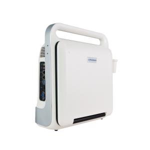
Modern Design 4D Color Doppler Diagnostic System Device long working hour battery
Portable Ultrasound Color Doppler Diagnostic Scanner
4D/3D optional,2D standard
The machine applications include abdomen, obstetrics, gynecology, blood vessels, small organs, urology,neonates and pediatrics. Includes scanning modes such as B / CFM / PDI / PW / CW / M and provides excellent resolution and sensitivity. Support convex array probe, linear array probe, convex probe, cavity probe, phased array probe, 4D probe. Integrated network ultrasound workstation, support for DICOM transmission. Using embedded computing system, the system is safe, stable and high-speed operation into one.The XF3800 is a value choice beyond your expectation!
• Flexible lifting panel • Trapezoidal imaging
• Reverse Tissues Harmonic Imaging
• 15-inch LCD display with up and down adjustment
• Optional 3D or 4D
• Compact fuselage, easy to move
• Two-level backlit keyboard, more convenient to operate
• lt can meet the needs of clinical diagnosis and improve the confidence of diagnosis.
• Broadband frequency shift harmonic imaging
• It can effectively reduce noise and improve contrast resolution
• Intelligent speckle noise suppression imaging Intelligent speckle noise suppression technology can intelligently identify different tissue information in different spatial dimensions, inhibit the display of speckle noise edge information, and make the image more exquisite
Main Functions:
Having a full digital beam forming technology Scanning mode: Convex array, lumen, high-frequency linear array, phased array;4D probe(Optional) Probe parameters * Convex: R60 (R50) Center frequency 3.5MHz (range: 2-6MHz) * Linear: Center Frequency 7.5MHz (Range: 5-12MHz) * High-frequency Linear : Center frequency 9MHz (range: 5-13MHz) * Cavity probe(TVS): center frequency 6.5MHz (range: 5-10MHz) * Micro-convex: center frequency 3.5MHz (range: 3-6MHz) * High-frequency micro-convex probe: the center frequency of 5MHz (range: 4-8MHz) * Phased array probe: 3 MHz (range: 2-5MHz) * 4D probe : Center frequency 3 MHz (range: 2-5MHz)
XF3800(4D) Portable full digital color doppler ultrasonic system
--Dynamic range: 0~120dB adjustable;
--Display mode: B,B/B,M,B/M,CFM,CMF/B,PDI,B/PW,CW etc mode;
--Application mode: abdomen, kidneys, urinary system, obstetrics, gynecology, pelvic, small organ, muscle tissue, organ, breast, heart and other 11 kinds of models;
--Depth display: ≥250mm;
--B/D triple-purpose: linear array: B/PW D; convex array: B/PW D;
--Pseudo color processing: 16 kinds of pseudo color encoding can optional;
--Gain adjusts: 8 segments TGC, B/M/D/C is independently adjustable; TGC curve can show and hide automatically;
--Image magnification: picture in picture zoom in and zoom part function;
--Self-motion optimize function: Built-in multiple check type, according to different inspection organs, preset best image check condition, reduce the adjusting operation keys;
--One-click optimization function: preset several parameters adjusting focus on a button, a key to realize image fast optimization;
--Image mode: digital beam forming, tissue harmonic imaging;
--IMT:Automatic measurement and analysis vasculer intima;
--Acoustic output: Mechanical index and thermal index real-time display;
--Acoustic power:Step is adjustable, real-time display;
--Gray scale: 256 scales;
Measurement and calculation:
--Distance, circumference, area, volume, angle, ratio, and stenos
rate.
--M mode routine measurement: Heart rate, time, distance, speed,
ratio, etc.
--Gynecology measurement: Uterus, cervix, endometrial, ovary,
follicular.
Obstetrics measurement:
EGA, ETD, fetal weight estimation, AFI index, OB report (including
OB tables).Cardiology measurement: LV measurement.
Urology measurement:
Prostate volume, displacement volume, bladder capacity, and
residual urine output.
PW measurements: Time, speed, Heart Rate, RI, PI, etc.
Other measurement:
Slice volume measurement, hip joint angle measurement.
Image storage: Image storage, video storage, cine loop, disk
storage capacity≥160G;
Patient data: Medical record management, report inquiry and
printing, image video output( HDD ,USB,Optional DVD-RW),built-in
ultrasound workstation;
Reporting system:
automatic report generation system, and can be full screen
characters in both Chinese and English editor;
Output interface:SR323,USB,DICOM interface;
PW MODE
| Measurement | Function |
| Time | Measure the time interval between any two points |
| Heart Rate | measure the time interval between 1 ~ 2 heartbeat cycle, calculate the heartbeat per minute |
| Speed | Measure the speed of a point on the doppler spectrum waveform and pressure difference |
| Acceleration | Measure the speed between two points and the time interval , computing speed difference and acceleration |
| Resistive Index | In waveform of PW spectrum image of blood flow, measure speed and pressure difference of the two peaks, calculate the resistance index and ratio value |
| Pulsation index | Arterial blood systolic peak (A or S) and valley value of end-diastolic (B or D), to calculate the S/D or A/B ratio, at the same time can also be calculated pulsation index (PI = A - B/mean value A, B) |
| Maximum differential pressure | The average pressure difference of maximum velocity wave spectrum |
| The average differential pressure | The average of the pressure gradient in the all recording area |
Main Technical Indexes:
The performance requirements of gray-scale imaging mode
The color ultrasonic at the gray-scale imaging performance mode should comply with the provisions of the
table 2.1
Table 2.1 At the Gray-scale imaging mode the performance of the probe
| Performance indexes | Probe type and nominal frequency | |||
| 2.0≤f<4.0 | 2.0≤f<5.0 | 5.0≤f<8.0 | 5.0≤f<10.0 | |
| a) probe type and model | phased array (type TP16A) | Convex array (type TC60A) | Cavity (type TC10A) | Linear array (type TL40A) |
| b) nominal frequency (MHz) | 3.0 | 3.5 | 6.5 | 7.5 |
| c) Scan depth(mm) | ≥140 | ≥160 | ≥40 | ≥50 |
| d) Lateral resolution (mm) | ≤3(depth≤80) ≤4(80 | ≤3(depth≤80) ≤4(80 | ≤2(depth≤30) | ≤2(depth≤40) |
| e) Axial resolution (mm) | ≤2(depth≤80) | ≤2(depth≤80) ≤3(80 | ≤1(depth≤40) | ≤1(depth≤50) |
| f) Blind area (mm) | ≤7 | ≤5 | ≤4 | ≤3 |
| g) Transverse geometry precision (%) | ≤20 | ≤15 | ≤10 | ≤10 |
| h) Longitudinal geometric location accuracy (%) | ≤10 | ≤10 | ≤5 | ≤5 |
| i) Slice thickness (mm) | ≤5 | ≤5 | ≤5 | ≤5 |
| j) Perimeter and area measured deviation (%) | ≤±20 | ≤±20 | ≤±20 | ≤±20 |
| k) M mode time display error (%) | ≤±10 | ≤±10 | ≤±10 | ≤±10 |
We are an experienced Manufacturer in China, focusing on research development and production in black
and white ultrasound and color doppler ultrasound scanner since 2005. We have been doing OEM and ODM more than hundreds of companies and factories over past 15 years.
Our products are affordable , reliable and competent. We sell over 10000 units to all over the world, and repair rate is less than 1%, all production process is strictly exectued with CE and ISO.