
Add to Cart
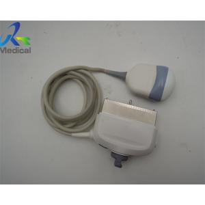
GE RAB4-8-D Wideband Curved Convex Array Used Ultrasound Probe Doctor Supplies Medical Scanner
1. Type: curved array
2. Frequency: 2-8 MHz
3. Compatible system: logiq e9/voluson e6/voluson e8
4. Application: abdominal, obstetrics, and gynecological
5. Condition: original, in good working condition
6. With 60 days warranty
| GE Ultrasound System |
| LOGIQ P5, LOGIQ P6, LOGIQ S7, LOGIQ S8, VOLUSON 730, VOLUSON 730 PRO, VOLUSON 730 EXPERT, VOLUSON S8, VOLUSON P8, VOLUSON S6, VOLUSON S20, VOLUSON P8, VOLUSON E10, VIVID E9, LOGIQ BOOK, LOGIQ 5, LOGIQ 7, LOGIQ 9, LOGIQ E9, LOGIQ F6, LOGIQ F8, VIVID I, VIVID Q, VOLUSON I, VOLUSON E, VIVID S5, VIVID 3, VIVID 7... |
Common ultrasonic Probe Damage(Convex, Linear, Sector, Endocavity probes)
| Common ultrasonic probe damage | Solutions |
| Lens damage, wear, holes, swelling, delamination | Lens replacement |
| Strain relief damage, separation | Strain replacement |
| Nosepiece and probe separation and cracks | Strain replacement |
| Cable cuts | Cable patches, possible cable replacement |
| Connector housing electrical damage | Major and minor electrical repairs, pin module replacement |
Knowledge Point
Color Doppler flow imaging
Color Doppler flow imaging system can simultaneously display B-type image and Doppler blood flow data (blood flow direction, flow velocity, flow dispersion) of dual ultrasound scanning system. Color power angio(CPA) detected the backscattered energy of blood cells in the blood flow, which did not distinguish the flow direction, and had nothing to do with the angle θ (the angle between the direction of sound wave and the direction of blood flow). CPA can improve the sensitivity of blood flow detection, especially suitable for displaying low-speed blood flow of small vessels, but can not show the direction of blood flow.
Harmonic imaging
Harmonic imaging exploits non-linear propagation of ultrasound through the body tissues. When the frequency of emitted sound wave is f 0, the frequency of echo (due to reflection or scattering) includes f 0 (called fundamental wave), 2f 0, 3F 0 and so on The second harmonic (2f 0) has the largest energy.
Ultrasonic harmonic imaging (UHI) is based on the information of human body carried by the second harmonic in echo (reflection or scattering). Harmonic imaging without UCA is called native harmonic imaging or tissue harmonic imaging. Harmonic imaging using UCA (ultrasound contrast agent) is called contrast harmonic imaging.
Daily and Long-Term Storage of ultrasound transducer
1. Always store transducers in the transducer holders on the side of your system or on a securely mounted wall rack when you are not using them.
2. Ensure the transducer holders are clean before storing transducers
3. When storing transducers, use the cable-management clips, if available, to secure the transducer cable
4. Avoid storing transducers in areas of temperature extremes or in direct sunlight.
5. Store transducers separately from other instruments to avoid inadvertent transducer damage.
6. Before storing transducers, make sure they are thoroughly dry.