Product Details
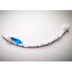
PVC Medical Grade murphy eyes with balloon 6.0mm Endotracheal Tube
Cuffed Nasal Endotracheal Tube
6.0mm Endotracheal Tube Cuffed Description:
HENAN AILE INDUSTRIAL CO., LTD is a company for operating medical
disposables ,our main products are specialized in anesthesia
products and respiratory products . In detailed, the anesthesia
products include Standard Endotracheal Tube, Preformed Oral/Nasal
Endotracheal Tube,Reinforced Endotracheal Tube.
Amoung them, the Standard Endotracheal Tube is a method of
inserting a special endotracheal tube into the trachea or bronchus
through the mouth or nasal cavity. And the Standard Endotracheal
Tube Cuffed is one of it, which has different size to adapt to
different medical needs, including size 3.0mm to 10.0mm.
Endotracheal Tube is a method of inserting a special endotracheal
tube into the trachea or bronchus through the mouth or nasal
cavity. And the Endotracheal Tube Cuffed is one type of it, which
has different size to adapt to different medical needs,including
3.0mm to 10.0mm.
A cuffed nasal endotracheal tube is a type of nasal tube used for
airway management and mechanical ventilation. It is similar to an
uncuffed nasal endotracheal tube, but it also includes an
inflatable cuff at the distal end of the tube.
Here are some important points about cuffed nasal endotracheal
tubes:
- Design: Cuffed nasal endotracheal tubes are typically made of
flexible plastic or silicone material. They have a beveled tip for
easier insertion through the nostril and advancement into the
trachea. The cuff is located near the distal end of the tube and is
inflated once the tube is in the proper position.
- Cuff inflation: The cuff of a cuffed nasal tube is inflated to
create an airtight seal within the trachea. This seal prevents air
leakage and reduces the risk of aspiration. The cuff is usually
inflated with air using a syringe, and the pressure is monitored to
ensure it remains within the recommended range.
- Advantages: Cuffed nasal endotracheal tubes offer several
advantages, including improved ventilation control, reduced risk of
aspiration, and the ability to provide positive pressure
ventilation. They are commonly used in adult patients, particularly
in critical care settings.
- Considerations: When using a cuffed nasal endotracheal tube, it's
important to carefully monitor the cuff pressure to avoid
overinflation or underinflation. Overinflation can lead to tracheal
damage, while underinflation may result in air leakage and
inadequate ventilation. Regular monitoring and adjustment of cuff
pressure are necessary to maintain an appropriate seal.
The specific steps for using a cuffed nasal endotracheal tube are
similar to those of other nasal intubation techniques, as described
earlier. However, additional attention should be given to proper
cuff inflation and monitoring to maintain a secure airway and
effective ventilation. Healthcare professionals trained in airway
management should follow established protocols and guidelines when
using cuffed nasal endotracheal tubes.
Product composition and function: | Size | 6.0mm |
| Balloon | Providing even pressure to maintain good sealing,reducing pressure
on the tissues of trachea |
| Radiopaque | Allowing clear identification of the tube on radiographic images |
| 15mm connector | Reliable connection to all standard equipment |
| Murphy Eye | Reducing the risk of occlusinon and maintaining airflow |
| Wire coil | Increasing flexibility, providing effective resistance to kinking |
| Valve | Ensuring continual cuff integrity |
- Smooth bevelled and carefully moulded hooded tip to assist
intubation and to provide high patient safety and comfort.
- The cuff of the endotracheal tube (ETT) is designed to provide a
seal within the airway, allowing airflow through the ETT but
preventing passage of air or fluids around the ETT.
- High volume/low pressure cuff helps to ensure an efficient low
pressure cuff seal, for intubation during long term ventilation.
- Intubation depth marks and pre-mounted 15 mm connector.
Application Features of 6.0mm Endotracheal Tube Cuffed :
- Suitable for both oral and nasal intubation.
- Tip-to-Tip X-ray line allows for safe positioning control.
- Murphy eye incorporated as an additional safety feature.
Intubation:
- During intubation, a physician usually stands at the head of the
bed looking towards the patient's feet and with the patient lying
flat. The positioning will vary depending on the setting and
whether the procedure is being done with an adult or child. With
children, a jaw thrust is often used.
- The endotracheal tube with the assistance of a lighted laryngoscope
(a Glidescope video laryngoscope is particularly helpful for people
who are obese or if a patient is immobilized with a suspected
injury to the cervical spine) is inserted through the mouth (or in
some cases, the nose) after moving the tongue out of the way.
- The scope is then carefully threaded down between the vocal cords
and into the lower trachea. When it's thought that the endotracheal
tube is in the proper location, the doctor will listen to the
patient's lungs and upper abdomen to make sure that the
endotracheal tube was not inadvertently inserted into the esophagus.
- Other signs that suggest the tube is in the proper position may
include seeing chest movement with ventilation and fogging in the
tube. When a doctor is reasonably sure the tube is in position, a
balloon cuff is inflated to keep the tube from moving out of place.
(In infants, a balloon may not be needed). The tube is then taped
to the patient's face.
Verifying Proper Placement:
- Once the tube is in place, it's important to verify that it is
truly in the proper location to ventilate the patient's lungs.
Improper positioning is particularly common in children, especially
children who have experienced trauma.
- In the field, paramedics have a device that allows them to
determine if the tube is in the correct position by a color
change.In the hospital setting, a chest X-ray is often done to
ensure good placement, though a 2016 review suggests that a chest
X-ray alone is inadequate, as is pulse oximetry and physical
examination.
- In addition to directly visualizing the endotracheal tube pass
between the vocal cords with a video laryngoscope, the authors of
the study recommended an end-tidal carbon dioxide detector
(capnography) in patient's that had good tissue perfusion, with
continued monitoring to make sure the tube does not become
displaced.
- In the setting of a cardiac arrest, they recommended using
ultrasound imaging or an esophageal detector device.
After the Procedure:
After the endotracheal tube is in place and a patient connected to
a ventilator, healthcare providers will continue to monitor the
tubing, settings, and provide breathing treatments and suctioning
as needed. Careful attention to oral care will also be provided.
Due to the location of the tube, patients who are conscious will be
unable to talk while the tube is in place.
Company Profile
We are a professional manufacture of Anesthesia and Respiratory
medical consumables !
Our factory is ISO certified Group which commit itself to R&D
of medical consumables. All products have been CE approved ,
involved in Anesthesia, Respiratory Series and are sold well in the
Middle East, Africa, Southeast Asia and the EU countries.
Meanwhile , we also have our own trading company , whose staff are
experts of medical industry and have more than ten years’
experience in this field. Therefore, we can provide “one-stop”
procurement service, saving your time and business cost . With our
assistance ,most of our clients have become the local well-known
distributor of all-round medical instruments. We sincerely hope to
establish a good business relation with you and devote a great
contribution to the development and growth of your company.
There are the CE Certificate pictures of our products: