
Add to Cart
non mydriatic fundus camera retinal imaging equipment advanced optical technology
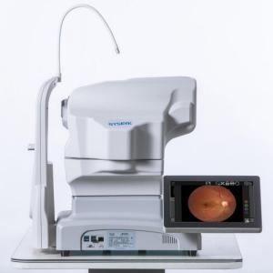
Automated Fundus Camera RETICAM 3100 is based on the traditional fundus camera of our company, after years of research and development, through binocular stereo vision and XYZ 3D image feature recognition automatic positioning, and through the division line to achieve automatic focus. Through CCD imaging feedback, the exposure light is measured and the exposure intensity is determined automatically and correctly. Focusing step is controlled according to the image resolution, and image definition is adjusted to obtain clear and accurate fundus image.Automated Fundus Camera RetiCam 3100 has excellent optical imaging capability, easy to operate, less dependence on the operator, easier to obtain images.Automated Fundus Camera RETICAM 3100 Fundus auto fluorescence (FAF), as a new retinal imaging technology, has been widely used in clinical medicine since its advent. It has been demonstrated that FAF can be used to diagnose and monitor many fundus diseases, including posterior uveitis, quickly and noninvasively. As a new application of non-invasive, non-contact, non-invasive, rapid and repeatable observation and monitoring of many fundus diseases including posterior uveitis, FAF has begun to play its unique role, and has attracted more and more attention of ophthalmologists and medical technicians.
specifications:
| Field of view | 50° |
| Field of view tolerance | ±7% |
| Resolution | |
| Center of view | ≥60 lp/mm |
| Field of view center (r/2) | ≥40 lp/mm |
| At the edge of the field of view (r) | ≥25 lp/mm |
| Magnification | 1.3 times |
| Required pupil diameter | 4.0 mm or more (3.3 mm or more when using the small pupil shooting function) |
| Working distance | 35 mm |
| Focus adjustment range | ±25D |
| Shooting light | Auto: with shooting mode |
| Manual: can be set manually | |
| light source | |
| Illumination light | Infrared led |
| Shooting light | Xenon lights |
| reflected light | |
| Scattered light | |
| camera | Digital camera |
| Fixing light | Internal fixing light (LED) |
| External fixation light | |
| Moving range | |
| Stage | 90 mm left and right, 35 mm front and rear |
| Main unit moves vertically | 30 mm |
| Chin-rest tray movement range | 60 mm |
| Rated power supply | AC 100V~240V,50/60Hz |
| size | 380 mm(L)x 550 mm(W)x 475 mm(H) |
| weight | About 26.5kg |
| When in maximum light intensity/hole bar state | |
| Spectral wavelength | 305 nm~1100 nm |
Company Introduction
Focusing on the two major public health problems of adolescent myopia and elderly ophthalmology, our company researches new diagnostic and treatment equipment for ophthalmology, develops low-cost applicable technology products, realizes industrialization, reduces the cost of social medical and health system, and serves the strategic needs of national health.The company always adhere to the "high-tech,new vision" for the enterprise development concept.We have a strong technical force, has been awarded 11 patents, and obtained 13,485 quality system certification.Bio has established an efficient marketing team and a perfect after-sales service system to provide medical equipment with the highest cost performance and meticulous service.