Add to Cart
Accurately Determine The Location Of Eye Lesions SD OCT Machine
SD OCT Machine examination mainly includes:
1. Macular hole:SD OCT Machine can clearly show the defect of retinal neuroepithelial light band in macular area.
2. Central serous choroidal retinopathy: SD OCT Machine appears as a neurodermal bulge in the macular area, beneath which fluid hyporeflectance is visible.
3. The anterior membrane of the retina: OCT is manifested as a highly reflective signal in the macular area close to the front of the retina, which can be continuous or intermittent
4. Polypoid choroidal vascular lesions: OCT can show the location, shape and range of detachment of retinal neuroepithelium and pigment epithelium, as well as high and steep detachment of pigment epithelium. The detachment lumen can be clear serous fluid or blood of different concentrations.
5. Age-related macular degeneration: OCT can clearly show vitreous warts, choroidal neovascularization and their size and shape, as well as macular sac.
6. Diabetic retinopathy: The etiology and course of the glycoreticulum are varied, but the typical fundus changes in each period can be shown in COT. Such as fundus punctate exudation, macular fovea detachment, hemorrhage, macular cystic edema and so on.
7. High myopia fundus lesions: OCT can show all retinal levels and choroid deletion in the nasal atrophic arc of optic disc, and know whether there is atrophy and thinning of the posterior polar retina, retinal cleavage and shallow retinal detachment, and vitreous macular traction.
8. Study on early diagnosis, follow-up and pathogenesis of glaucoma: frequency-domain OCT has potential advantages in the diagnosis and follow-up of glaucoma. It can show different degree of thinning of nerve fiber layer around optic disc, thinning of optic nerve fiber layer, glaucoma cup and other pathological changes that can not be found or difficult to be found by conventional fundus examination.
9. Keratopathy: The anterior segment mode of OCT can measure the thickness of the cornea, display the infiltration and thinning caused by corneal inflammation, corneal foreign body and the depth of foreign body, etc., which is of great significance for the diagnosis and further treatment of keratopathy.
10. Angle examination: The Angle mode of OCT can distinguish open Angle glaucoma from Angle closure glaucoma. Compared with UBM examination, OCT does not touch the cornea and is easy to operate, which can partially replace UBM in the diagnosis and treatment of glaucoma.
Specifications of SD OCT Machine
| Methodology | Spectral domain OCT |
| Axial resolution | ≤6 µm (in tissue) |
| Transverse resolution | ≤20 µm (in tissue) |
| Scan depth | ≥2.5 mm (in air) |
| Scan range | ≥6 mm |
| Scan speed | ≥24,000 A-scans/sec, up to 36,000 A-scans/sec |
| Scan modes | 3D, Raster, Circle |
| Fundus image | OCT en face |
| Focus adjustment | -15D to +15D |
| Pupil diameter | ≥3 mm |
| OCT light source | 840 nm SLD |
| Optical power | 750 µW (at cornea) |
| Operation | 13.3” touch screen, optional external mouse or keyboard 100-240 V, 50/60 Hz |
| Power supply | 497 mm × 395 mm × 490 mm (L × W × H) |
| Dimensions Weight | 34 kg (75 lbs) |
Compact Design
Features:
1. Compact Design: Everything is inside this compact body. No
external computer is needed. Plug in the power cable, then you are
ready to go.
2. PC Inside: Data acquisition and processing are accomplished by
the internal computer. The data can be transported via ethernet or
external harddrive. Peripheral devices, such as keyboard or
printer, can be connected to the computer ports.
3. Easy Installation:No complex connection or setup. Compact body
can fit in even small space.
SD OCT Machine has never been so affordable. More people can benefit from the advanced technology now.
Step 1
Choose left or right eye. Push the direction buttons to move the
target eye to the center of the field of view, then start data
acquisition.
Step 2
Enter the data acquisition interface. The device is able to search
for the OCT signal and optimize it automatically.
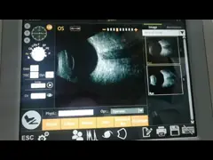
Step 3
Browse and analyze the acquired images.
Company Introduction
Focusing on the two major public health problems of adolescent myopia and elderly ophthalmology, our company researches new diagnostic and treatment equipment for ophthalmology, develops low-cost applicable technology products, realizes industrialization, reduces the cost of social medical and health system, and serves the strategic needs of national health.The company always adhere to the "high-tech,new vision" for the enterprise development concept.We have a strong technical force, has been awarded 11 patents, and obtained 13,485 quality system certification.Bio has established an efficient marketing team and a perfect after-sales service system to provide medical equipment with the highest cost performance and meticulous service.