15 Inch Multiple Beam Synthesis Veterinary Ultrasound Machine For Small Animals
Brand Name:Golead
Certification:ISO13485
Model Number:P60V
Minimum Order Quantity:1 Unit
Delivery Time:2 working days
Payment Terms:T/T
Contact Now
Add to Cart
Verified Supplier
Location:
Chengdu Sichuan China
Address:
03/03/01, No.366, North Hupan Road, New Tianfu Zone, China(Sichuan) Free Trade Area, Chengdu, China.
Supplier`s last login times:
within 1 hours
Shipping
lt's easy to get a shipping quote! Just click the button below and complete the short form.
Get Shipping Quote
Product Details
Company Profile
Product Details
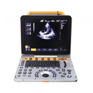
15 inch Portable Vet Ultrasound Machine for Small Animals
Overview
Portable veterinary ultrasound machine P60V used advanced and
stable PC system, combing excellent images, all-round functions,
rich image management software together to meet the needs of
various animal groups in different environments. The built-in
battery with large capacity imported chip, dual probe interface,
canbe configured with a variety of probes to satisfy the needs of
different examination modes
Parameters
| 1. Product Name: 15 inch Portable Vet Ultrasound Machine for Small Animals |
| 1.1. Structure Style: Portable |
2. Application |
3. System Technical Specifications and Summary |
| 3.2. Digital Beam Enhancer |
| 3.3. Multiple Beam Synthesis |
| 3.4. Full-Digital 2D Gray Scale Imaging |
| 3.5. Tissue Harmonic Imaging (THI) |
| 3.6. B/C Real-time Two Synchronous Imaging |
| 3.7. M Mode Imaging |
| 3.8. Anatomic M Mode Imaging (Sampling line≥3) |
| 3.9. Color Doppler Imaging (CFM, PDI, DPDI) |
| 3.10. Spectral Doppler Imaging (PW, HPRF PW, CW) |
| 3.11. Tissue Doppler Imaging (TVI, M-mode, Spectral Imaging, etc.) |
| 3.12. Four-dimensional Supersonic Image Formation |
| 3.13.★Contrast Tuned Imaging (optional) |
| 3.14.Wide Field Imaging (optional) |
| 3.15. Space compound imaging (application to Abdominal, GYN, vessel, superficial small organs, can be dual image contrast display at same time) |
| 3.16. Frequency & Focus Compound |
| 3.17. Extended field-of-view (EFOV) |
| 3.18. Real-time dual image contrast display |
| 3.19. B/C/D Real-time three synchronous imaging |
| 3.20. Speckle Reduce Imaging (SRI) |
| 3.21. Elastography (optional) |
| 4.★Operation Interface: support 10 different languages |
5. System Specification |
|
5.3. 2D Imaging Mode |
5.4. Color Doppler Imaging Mode |
5.5. Spectral Doppler Imaging |
5.6. ★PView wide field imaging (optional) |
5.7. Probe: wideband with frequency conversion, independent
frequency conversion under B and CFM |
5.8. Technical requirements for electronic convex probe: |
5.9. Technical requirements for electronic linear probe: |
6. Measurement / Analysis and Reports |
6.2. Specialized measurement:
bile duct, pancreas, spleen, abdomen aortic diameter, kidney. |
7. Peripherals Section |
8. Cine Loop and Original Data Processing |
9. After-Sale Service |
Applications
Bovine, Equine, Ovine, Swine, etc
Standard configuration
Veterinary Ultrasound Machine with Mirco-convex Probe and linear
probe
Optional
Covex probe
Rectal linear probe
Phased array probe
Image Gallery

15 Inch Multiple Beam Synthesis Veterinary Ultrasound Machine For Small Animals
Inquiry Cart
0