Add to Cart
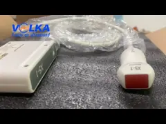
Alpinion E-Cube 8 original convex array ultrasound probe C1-6CT
Form Factor: Convex Array
Applications: OB/GYN,Abdominal
Frequency Range: 1.0 - 6.0 MHz.
Compatibility: E-Cube 5, E-Cube 8
More details welcome to contact with us!
Transducers powered by PowerView™: C1-6CT / P1-5CT
The PowerView™ technology is applied to the E-CUBE 8‘s Convex and
Phased array transducers. The PowerView™ technology disperses heat
generated by the transducers, improving the E-CUBE 8‘s durability
and ensuring the stability of each diagnosis. The increased
efficiency of ultrasonic waves enhances the signal sensitivity and
improves the expression of clinical images. Integrated with
Alpinion‘s innovative technologies, the E-CUBE 8 promises superior
image resolution and penetration with a reasonable price.
High performance linear transducers: L3-12H / L3-12HWD
The high-density linear transducers can be attached to the E-CUBE
8. Several footprint width options and high-quality linear images
help with breast/thyroid/musculoskeletal/ vascular examinations.
Use of a flagship model-grade platform
Equipped with Alpinion’s top model-grade platform, the E-CUBE 8 has
high end hardware and software. The resolution, contrast, and
uniformity of 2D images have been improved, and with the addition
of the Dual pulser, clear and accurate Doppler data can be
displayed while maintaining sharp 2D images in the Doppler Combined
Mode.
Optimal Imaging Suite™ Plus
By combining Alpinion‘s image optimization processing technologies:
SCI, FCI, FTHI, PITHI, and SRI/FullSRI™, artifacts are eliminated
effectively and boundaries between tissues are distinguished more
clearly. Furthermore, a broader grayscale range enables the
expression of richer tissue textures and accurate data.
Xpeed™
Simply press the Xpeed™ button once to quickly optimize images in
2D Mode and Spectrum Doppler Mode. Detect, predict, and adjust the
Dynamic range level in real-time. It displays images optimized and
customized for different clinical cases.
SSD for quick exam preparation
The E-CUBE 8 uses high-end hardware, including an SSD. These
enhance stability when using the system and the fast boot time
makes speedy preparation for examination possible.
USB 3.0 for better patient care
The E-CUBE 8 uses a USB 3.0 port. Compared to current USB 2.0
ports, the data transfer speed for USB 3.0 ports is about ten times
faster. The USB 3.0 port reduces the transfer time when exporting
data for patients or research, allowing the user to focus more on
patient care.
21.5-inch full HD LED monitor
The 1,920×1,080 pixel high-resolution monitor delivers sharp, clear
ultrasound images. With the use of IPS (In-Plane Switching)
technology, image distortions do not occur and a wider field of
view is provided. As the user can review images easily without
being restricted by location or environment, the accuracy and
convenience of each diagnosis is improved.
10.4-inch touchscreen
By applying an intuitive UI design to a capacitive touchscreen with
high sensitivity, like the one used on tablet computers, the
convenience and speed of using the touchscreen have been improved.
Power Preset
The user can load a system preset saved in advance with a single
touch. Quick and easy application of presets will shorten the image
setup time.
User-friendly control panel
The E-CUBE 8‘s control panel keys are arranged in the most
efficient and intuitive manner for examination. Frequently used
functions can be assigned to the three user keys, which are
arranged for easy access on the control panel. By minimizing the
number of unnecessary keypresses, the E-CUBE 8 reduces user fatigue
and increases the operating speed. The brightness level of the
backlight of the control panel is adjustable, enabling it to be
used in a darker environment.
Easy-to-use keyboard
The E-CUBE 8 has a keyboard on top of the control panel, making it
easy to access. When the user needs to type text during an
examination, they can access the keyboard right away, reducing
unnecessary tasks and shortening the examination time.
Battery that frees you from space restrictions
The combination of compact exterior design and attached battery
makes the E-CUBE 8 much easier to transport. The user can move to a
different location while in Exam Mode without connecting the power
cable and resume the examination straight away. More time can be
reserved for patient care by reducing the time spent on turning the
system back on.
Gel warmer developed for patient convenience (optional)
The E-CUBE 8‘s gel warmer warms up the ultrasound gel before
examination. The temperature can be adjusted in three steps
according to examination circumstances. This will help provide
patients with a positive examination experience.
The E-CUBE 8 aims to create a user and patient-oriented design and workflow. The user can better focus on patient care, as the E-CUBE 8 can be used easily and conveniently in different clinical environments.
The E-CUBE 8 is a multi-purpose system that can be used in all specialized areas that require ultrasound imaging such as internal medicine, obstetrics/gynecology, orthopedics, etc. It broadens the application range of ultrasound examination and ensures accurate diagnosis using premium-grade software diagnostic tools.
Auto IMT
When the user draws a line in the area where the carotid intima
media thickness is to be measured, the thickness will be measured
automatically and displayed on the screen. Measurements can be made
more quickly and accurately down to the millimeter level,
regardless of the user‘s proficiency.
CUBE Strain™
This is a non-invasive examination method that is used to assess
the myocardial function more objectively. The user can track
speckles in 2D heart images, digitize the movement of each
myocardial segment, and check quantified data.
Stress Echo
The optimized examination workflow allows the user to perform a
Stress Echocardiogram more quickly and conveniently, aiding early
diagnosis of chronic coronary heart disease.
Live HQ™
With the improved volume rendering technology, the light source can
now be moved freely and the optimized color map can be applied in a
variety of different ways. Realistic volume images make fetal
anatomy easier to understand, which leads to more accurate and
quicker diagnosis, and helps create a bond between the mother and
the unborn baby.
Auto NT
When the user draws a ROI box in a desired measurement area during
a nuchal translucency scan, the maximum thickness will be
automatically measured and displayed on the screen. Examination
results can be checked quickly in busy examination environments.
Volume Master™
Volume Master™, Alpinion’s 3D/4D features, enables you to obtain
reproducible planes and better anatomical views which are not
obtainable with 2D scanning. Multi Planar Rendering (MPR), Cubic
View, and Multi Slice View (MSV) provide the clinical benefits of
CT or MRI.
Volume Advance™
On top of Volume Master™, Volume Advance™ provides the following
more advanced features for handling volume data: Free Angle MSV,
AnySlice™, and Volume Analysis. You can slice a desired section and
display slices consecutively. Therefore, anatomical and
pathological characteristics and volume information can be
delivered more accurately and in detail.
Elastography
Elastography intuitively shows the relative differences in tissue
elasticity caused by external pressure by using colors. It provides
additional pathological information and helps reduce the need for
unnecessary biopsies. The Indication bar shows whether the amount
of pressure on tissues is appropriate in real-time on a scale of 1
to 6, adding to the credibility of results.
Needle Vision™ Plus
Using Beam Steering technology, this feature is useful in showing
the shape and orientation of the needle. During invasive
ultrasound- guided procedures using the linear transducer, the
needle can be viewed more clearly by adjusting the beam angle in
three steps, ensuring more accurate and safer procedures.
Alpinion develops and manufactures transducers in-house.
Reliable quality / Best compatibility / Cheaper maintenance /
Faster repair
Transducer Guide
Convex
C1-6CT
C-Architecture
(PowerView™) Convex
OB, GYN, Abdomen, Urology, Pediatric, Musculoskeletal (MSK),
Vascular, Emergency Medicine (EM)
C5-8NT
Micro Convex
Pediatric, Abdomen, TCD (Transcranial), Cardiac, Vascular, OB,
Emergency Medicine (EM)
Volume Convex
VC1-6T
Volume Convex
OB, GYN, Abdomen, Urology, Pediatric, Emergency Medicine (EM)
Linear
L3-12H
High Density Linear
Small Parts, Musculoskeletal (MSK), Vascular, Abdomen, Pediatric,
TCD (Transcranial), Emergency Medicine (EM)
L3-12HWD
High Density Linear, 64mm
wide footprint
Small Parts, Musculoskeletal (MSK), Vascular, OB, Abdomen,
Pediatric, Emergency Medicine (EM)
L3-12T
Linear
Small Parts, Musculoskeletal (MSK), Vascular, Abdomen, Pediatric,
TCD (Transcranial), Emergency Medicine (EM)
Phased Array
P1-5CT
C-Architecture (PowerView™)
Phased Array
Cardiac, Abdomen, Pediatric, TCD (Transcranial), Vascular, OB, GYN,
Emergency Medicine (EM)
SP3-8T
Single Crystal Phased Array
Pediatric, Cardiac, Abdomen, TCD (Transcranial), OB, Emergency
Medicine (EM)
Endocavity
EC3-10T
Endocavity (Straight)
Urology, GYN, OB, Transrectal,Transvaginal, Vascular, Emergency
Medicine (EM)
EV3-10T *
Endocavity (Curved) GYN, OB, Urology, Transrectal, Transvaginal,
Vascular, Emergency Medicine (EM)
E3-10
Endocavity (Straight)
GYN, OB, Urology, Transrectal, Transvaginal, Vascular, Emergency
Medicine (EM)
Pencil
CW5.0
Pencil Typed
Cardiac, Vascular
CW2.0
Pencil Typed
Cardiac