Add to Cart
High precision tungsten devices for CT collimators and detectors
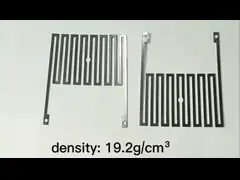
1. X-Ray Tube Side Collimator:
mounted in front of the CT bulb, is the first component through which the X-rays pass. The X-rays coming from the bulb are conical and pass through a tungsten device with slits to obtain a thin sheet-shaped beam [1], whose main function is to perform layer thickness selection of the X-rays by controlling the width of the X-rays beam in the direction parallel to the long axis of the body and thus controlling the thickness of the scanned layer.
2. Detector side collimator :
Its slits are individually aligned with each detector so that the detector receives only rays incident perpendicular to the detector, minimizing interference from scattered rays in other directions.
Working Principle
The detector is a device that converts the X-ray signal into an electrical signal after absorption and attenuation by the human tissue, and there are two types of detectors, fixed and gas. Both types of detectors are used in modern CT devices, and the choice of which measurement system to use depends on which characteristics are favored. Gas detectors use the principle of ionization of the inert gas xenon (Xe) gas, where the incident X-rays ionize the gas and the intensity of the incident X-rays is measured by measuring the magnitude of the current. The gas detector uses a tungsten device for both the spacer and the collection electrode. The spacer is in the same direction as the incident X-rays and acts as a back collimator, which prevents the scattered rays generated by the measured body from entering the ionization chamber. When a beam of X-rays enters the high-pressure xenon gas chamber, the xenon gas ionizes and the positive ions move to the negative electrode, while the negative ions are collected by the collecting electrode (tungsten device), generating a signal current, which directly reflects the amount of X-rays absorbed by the detector.