Product Details
P60 Color Doppler Diagnostic Ultrasound System
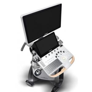
Size and Weight
Width: approx. 550 mm
Depth: approx. 740 mm
Height: approx. 1360 mm (the height is
measured when the control panel and the
monitor are adjusted to the lowest position)
Weight: approx. 84 Kg
Monitor
Medical high resolution monitor
Resolution: 1920*1080
Viewing angle: 178°(horizontal), 178°(vertical)
Swivel angle: ±40°
Up/down angle: -90° to 25°
Contrast and brightness: 0 - 100 adjustable
Monitor Arm
-- Swivel angle between upper arm and lower
arm: ≥ 100°, horizontal
-- Swivel angle between lower arm and control
panel: ≥ 50°, horizontal
-- Extend direction: up/down and left/right
Touch Screen
Medical high resolution monitor
Resolution ratio: 1920×1080
Viewing angle: 160° (horizontal), 160°
(vertical)
Inclination angle: adjustable
User-defined parameter preset layout
Control Panel
User-oriented design
Backlight design: panel buttons
Multiple defined-keys
TGC: 8 levels slider controls
Trackball sensitivity: adjustable
Adjustable height range: 0 - 230mm
Full-sized backlit keyboard on the panel
| Hardware: |
| P60 Main Unit |
| 21.5" High Resolution LED Color Monitor |
| 13.3" High Resolution Touch Screen |
| Height Adjustable and Rotatable Operation Panel |
| Five Active Probe Ports |
| One Pencil Probe Port |
| Build-in ECG Module (Including Hardware and Software) |
| Wifi Module |
| Hard Disk 1T |
| Software: |
| B (2B & 4B) Mode |
| M Mode |
| Anatomic M Mode |
| Color M Mode |
| Color Doppler Flow Imaging |
| Power Doppler Imaging / Directional Power Doppler Imaging |
| Tissues Doppler Imaging |
| Pulse Wave Doppler Imaging |
| Continuous Wave Doppler Imaging |
| High Pulse Repeat Frequency |
| Tissue Harmonic Imaging |
| Pure Inversion Harmonic Imaging |
| Compound Imaging |
| Tissue Specific Imaging |
| Image Rotation |
| μ-Scan: Speckle Reduction Technology |
| SR Flow |
| Simultaneous Mode (Triplex) |
| FreeHand 3D Imaging |
| B Mode Panoramic Imaging / Color Panoramic Imaging |
| Lateral Gain Compensation |
| Trapezoid Imaging |
| Widescan Imaging |
| Biopsy Guide |
| C-xlasto (Strain Elastography) |
| Needle Visualization Enhancement (VIS-Needle) |
| Bladder Volume Measurement |
| Zoom (Pan-Zoom / HD-Zoom / Scr-Zoom) |
| TEI Index |
| PW Auto Trace |
| Auto IMT |
| Auto EF |
| Auto OB: BPD / HC / AC / FL / NT / HL |
| S-Guide |
| Build-in User Mannual (Help) |
| Sono-help (Scanning Tutorial) |
| DICOM 3.0: Store / C-Store / Worklist / MPPS / Print / SR / Q&R |
Company Profile
Shenzhen Kenid Medical Devices CO.,LTD
established in 2010,is a leading manufacturer which specialized in
producing medical dry films,we have self-owned brand "KENID".
Our company insists management strategy which"professional"with"the
famous brand",with deeper promotion of the products,we have own
many superior customer not only from domestic but also
international market.
Our products have the following advangtage:
1、High speed with high quality
Using top dry imaging technology"direct digital imaging
technology",to provide users with high density,high contrast
images,make the picture more perfect.Dry technology needn't potions
flush or dark room equipment,so that users can print high quality
diagnosis images more easily and quickly.
2、Environmental friendly products
"direct digital imaging technology"take more facility to
users,needn't potions flush or dark room equipment,for you to
dispense dirty and tedious darkroom equipment debugging.
3、Stable clearer image.
Kenid medical films are using kind of organic,thermal inductance
emulsion membrane,then be protected by special protection layer to
avoid scratch and moisture.Top production process ensure
stability,high quality images with high contrast,low film fog.With
non-photosensitivity,"KENID" medical films provide more
conveniency,improved efficiency,reduced errors.
"KENID"people thank you for your support,trust and help.We will,as
always,adhere to the "innovation as power,to the quality of
survival"development path,focus on medical image field,provide
better products,superior serviceDD