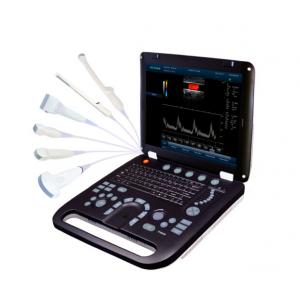
4D Color Doppler Ultrasound Scanner with 3.5MHz / R40 Volume Probe
Brand Name:Huge
Certification:CE
Model Number:EC-50D
Minimum Order Quantity:1
Delivery Time:3-5 working days after get payment
Payment Terms:MoneyGram, Western Union, T/T,paypal
Contact Now
Add to Cart
Active Member
Location:
Shenzhen Guangdong China
Address:
7A,Tongshengkeji Building, No.3 Huahui Road, Dalang street, Longhua new district, Shenzhen City, China. Zipcode: 518109
Supplier`s last login times:
within 48 hours
Shipping
lt's easy to get a shipping quote! Just click the button below and complete the short form.
Get Shipping Quote
Product Details
Company Profile
Product Details
4D Color Doppler Ultrasound Scanner with 3.5MHz / R40 Volume Probe
Quick Detail:
Color Doppler (CFM)
Power Doppler (PDI)
Directional Power Doppler (DPDI)
Pulsed Wave Doppler (PWD)
continuous wave doppler CW
B+PWD (Duplex)
B+CFM/PDI/DPDI+PWD (Triplex)
High Pulse Repetition Frequency (HPRF)
Tissue Harmonic Imaging (THI)
Description:
Ultrasound diagnosis technique is to use the ultrasonic as information carrier, the ultrasonic probe transmits ultrasonic to human body, and through the same probe (or called as transducer)receive echo with information on human body tissue, then through information extraction and processing to realize the inspection and diagnosis on human body tissue, it has some advantages in safety, no wound, direct viewing, real time, repeatable inspection, convenient operation, wide application, inexpensive price and stronger discriminability to parenchyma. It has taken possession of very important position at the current four image diagnosis techniques in medicine, it has been widely applied to clinic diagnosis, family planning and getting well and health protection, the ultrasonic diagnostic instrument has become a popular and conventional diagnosis system.
Applications:
- Abdomen section (adults and pediatrics) : to detect abnormalities from the structure images of liver, kidney, pancreatic gland, gall bladder, spleen, gastrointestinal tract and urogenital organ.
- Abdomen section (obstetrics and gynecology): to detect and discover abnormalities from the structure images of fetus, womb and pelvis so as to estimate the fetal age and weight and evaluate the fetal heart function.
- Chest section (adult and pediatric): to analyze M-mode image so as to detect abnormalities of the heart structure and function.
- Skin section (organella):to detect abnormalities from the rough evaluation of images of breasts, thyroid, and testicles, etc.
- Skin section (peripheral vessel): to detect and evaluate the vascular stenosis and occlusion from the images of peripheral vessels and flow measurement.
- Neonatal head section: to detect abnormalities of the cerebral volume and structure from the image of neonatal head.
- Urological section: to detect the left/right kidney, bladder and calculate residual urine volume, etc.
- Musculoskeletal section: to detect and discover abnormalities from the structure images of fetus and musculoskeletal in the application of obstetric and gynecology.
- Transvaginal section: to detect and discover abnormalities from the structure images of uterus, ovary, ovarian follicular, etc.
Specification:
Standard Configuration
1. EC-50D main unit
2. 3.5MHz/R40 volume probe
3. 4D imaging software package
4. phased array probe
5. 250ml coupling gel
6. AC adapter
7. Built-in rechargeable lithium battery
8. Protection bag
9. Grounding cable
10.Fuse (2)
11. Operation manual
Image:
B, B|B, 4B, B|M, M,4D
Color Doppler (CFM)
Power Doppler (PDI)
Directional Power Doppler (DPDI)
Pulsed Wave Doppler (PWD)
continuous wave doppler CW
B+PWD (Duplex)
B+CFM/PDI/DPDI+PWD (Triplex)
High Pulse Repetition Frequency (HPRF)
Tissue Harmonic Imaging (THI)

4D Color Doppler Ultrasound Scanner with 3.5MHz / R40 Volume Probe
Inquiry Cart
0