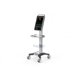
Ultrasound Scanner Equipment Color Doppler Machine With 2D Software For Human Use
Brand Name:BIO
Model Number:5000M
Minimum Order Quantity:1 unit
Delivery Time:7 working - days since getting full payment
Payment Terms:Credit card, Paypal, T/T
Type:Color Doppler Ultrasound Equipments
Contact Now
Add to Cart
Verified Supplier
Location:
Wuxi Jiangsu China
Address:
Room 408, No.801 Hongqiao road, Wuxi, Jiangsu, China
Supplier`s last login times:
within 39 hours
Shipping
lt's easy to get a shipping quote! Just click the button below and complete the short form.
Get Shipping Quote
Product Details
Company Profile
Product Details
Ultrasound Scanner Equipment Color Doppler Machine With 2D Software For Human Use
Function
-1 probe connector, which can be expand to 3 connectors by the probe expansion module
-18.5"high-resolution color LCD monitor
-Equipped with more than 4 beamformer function
-Equipped with THI (Tissue Harmonic Imaging), and second digital THI function
-Equipped with TSI (Tissue Specific Imaging) function
-Frequency compound imaging: adjustable under 2D and M modes
-Space compound imaging: adjustable under 2D and M modes with more than 3 levels
"-The scanning modes include 2D (2D ultrasound scanning diagnostic method), M (time motion, M mode diagnostic method), PW
(Pulsed Wave Doppler), CFM (Color Flow Mapping), PDI (Power Doppler Imaging)."
-Different frequencies are selected for 2D image and color image
-Equipped with biopsy guide line, guide by convex probe, transvaginal and linear probe
"-Equipped with bidirectional cine-loop function, gray scale cine-loop no less than 1024 frames, PW cine-loop time is no less than
100 seconds. Automatic/manual playback. The speed of the playback can be adjusted"
-Massive image storage (related with configured hard disk, no less than 500G)
-Equipped with 123 types of body marks; the probe position and scanning direction can be shown with arrow
Optional transducer
| Mode | Probe | Frequency |
| CR60 | Convex probe | 2-5Mhz |
| CR11 | Micro-convex probe | 5-8Mhz |
| CR20 | Micro-convex probe | 2-4Mhz |
| L25 | Linear probe | 5-12Mhz |
| L40 | Linear probe | 5-10Mhz |
| VR10 | Transvaginal probe | 5-8Mhz |
Specification
| Monitor | 18.5" LCD touch monitor |
| Video Output | PAL-D, S-video, NTSC, VGA, DVI |
| Digital Scan Converter | 628× 440 × 24 bits |
| Body Mark | 123 body marks with probe location |
| Probe Frequency | 2. 0MHz~10. 0MHz |
| Gray Scale | 256 |
| Color Scale | 24 |
| Frame Rate | Max. up to 70 f/s |
| Scanning Area | ≥ 320 mm |
| Density of Scanning Lines | Max 256 line/frame |
| Cine loop | ≥1024 frames |
| Biopsy | Optional biopsy guide |
| Display Mode | B, 2B, 4B, M, B+M, CFM, PDI, B+PW, B+CFM+PW, B+PDI+PW |
| Type of Focusing | Dynamic focusing, acoustic lens focusing, launching multipoint focusing |
| Scanning Technique | Dynamic apodization, dynamic aperture, dynamic frequency scanning, multi-acoustic beam |
| Pre-processing | low noise preamplifier, TGC, filtering, frame average, line average |
| Post-processing | Gamma correction, histogram, digital scan conversion (DSC), edge enhancement, noise rejection, smooth, ePure, grey scale transformation, pseudo-color, color persistence etc |
| Display Control | Freeze/Unfreeze, Left-right reverse, Up-down reverse, Polarity reverse, picture rotation (90°/180°/270°), color reverse, inverse-frequency spectrum, pseudo-color. |
| Control of Transformation and Adjustment on Sound Field | output acoustic power, PRF, focus position, scanning angle, image frame frequency, impulse duration, depth, sampling area dimension |
| General Measurement | 2D/CFM: Distance, area, volume (ellipse method), angle; M: Distance, time, slope, heart rate, simplified left ventricle function, complete left ventricle function; PW: Distance, max ventricular gradient, average ventricular gradient, time, S/D ration, blood flow rate, acceleration of blood flow, heart rate, pulsate & drag index. |
| Image Memory | Hard disk for massive image storage, min. 10,000 images permanent stored |
| Character | Date, time, patient name, device name, user name etc, user-defined annotation table, arrowhead and body marks |
| Measurement Software | Software packages of Abdomen, Urology, Cardiology, Obstetrics, Gynecology, Superficial Parts, Blood Vessels, Fetal Heart, Transcranial Doppler and Orthopedic Surgery; directly form diagnostic report based on measurement results. |
| Input Power | 120VA |
| Continuous Working Time | >8 h |
| Size(3 cartons) | 65*42*18cm;140*15*9cm;73*60*39cm |
| Weight | 40kg |

Ultrasound Scanner Equipment Color Doppler Machine With 2D Software For Human Use
Inquiry Cart
0